Diagnostics & Technology
A three-dimensional view of teeth, jaws, airway, sinuses and nerves.
Detail that standard X-rays cannot capture, and that surgical planning often depends on.
Request an Appointment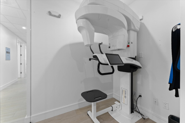
Detailed. Three-Dimensional. Explained to You.
What a Cone Beam Scan Shows
Cone Beam CT scanning is an advanced imaging technology that provides highly detailed, three-dimensional views of the teeth, jaws, airway, sinuses, nerves, and surrounding facial structures. This level of imaging is particularly valuable in oral and maxillofacial surgery, where treatment often depends on a detailed understanding of complex anatomy.
Where We Use It
At South Florida Institute of Oral Surgery, Cone Beam CT scans are used to support diagnosis and planning for a wide variety of procedures, including wisdom teeth removal, implant placement, TMJ evaluation, corrective jaw surgery, and more. The technology allows our surgeons to see what cannot always be captured with standard X-rays alone.
Accuracy and Patient Understanding
By giving us a more complete picture of the patient’s anatomy, Cone Beam CT imaging helps improve accuracy, reduce uncertainty, and support more predictable surgical planning. It also allows for better patient education, since we can visually explain findings and treatment recommendations in a more meaningful way.
Safety, Precision, Outcomes
This technology is one of the many ways South Florida Institute of Oral Surgery brings advanced care to every patient experience. Our goal is to use the most effective tools available to improve safety, precision, and outcomes.